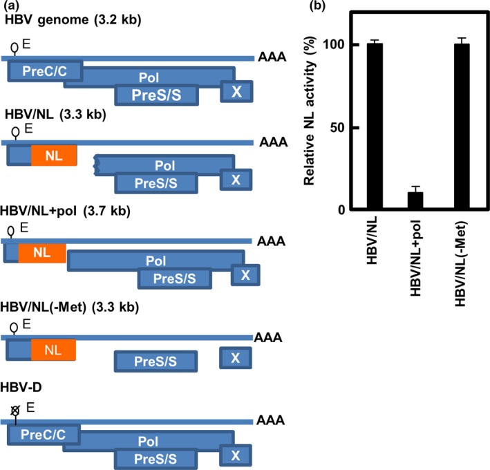
Figure 1.

Pregenomic structure of the reporter hepatitis B viruses (HBVs) and relative NanoLuc (NL) activity in cells infected by the viruses. (a) Pregenomes of wild‐type and reporter HBV, and putative ORFs of the virus are shown. The indicated sizes (kb) are of the pregenomes. A stretch of “A”s indicates a poly A tail of the putative pregenomic RNA. A lariat rope with “E” indicates an encapsidation signal and “X” on that indicates defect of encapsidation. The NL gene is inserted into the genome so as to be translated from its own initiator methionine. Virus was produced by co‐transfecting with the pUC1.2xHBV/NL, pUC1.2xHBV/NL+pol, or pUC1.2xHBV/NL(‐Met) and pUCxHBV‐D into HepG2 or HuH7 cells and the same volume of virus fractions was infected into PXB cells. (b) PXB cells were harvested 7 days after infection and NL activity in cell lysates was measured. The NL activity in the cell lysates was quantified (mean ± SD; n = 3). The data shown are of three independent experiments.
