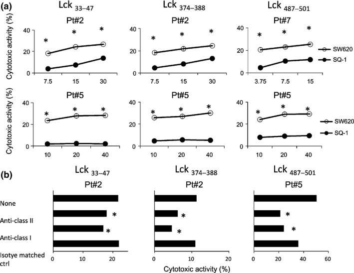
Figure 2.

Cytotoxicity of peptide‐stimulated peripheral blood mononuclear cells (PBMC). (a) PBMC from the three HLA‐A2+ cancer patients were stimulated in vitro with each of the Lck33–47, Lck374–388 and Lck487–501 peptides followed by testing of their cytotoxicity against HLA‐A2+ Lck+ SW620 and HLA‐A2− Lck+ SQ‐1 cells by a 6‐h 51Cr release assay at three different effector to target cell ratios. Representative results are shown in the figure. *P < 0.05 (statistically significant). (b) To determine the HLA class I‐restricted or class II‐restricted manner, 10 μg/mL of either anti‐HLA‐class I (W6/32: mouse IgG2a), anti‐HLA‐DR (L243: mouse IgG2a) or anti‐CD14 (H14: mouse IgG2a) mAb, as an irrelevant control, were added into wells at the initiation of the culture. Representative results are shown in the figure. *P < 0.05 (statistically significant). The genotypes of Pt.2, Pt.5 and Pt.7 were HLA‐DR04:05/13:02 and HLA‐A02:06/33:03, HLA‐DR04:03/09:01 and HLA‐A02:01/02:06, and HLA‐DR09:01/14:05 and HLA‐A02:01/31:01, respectively.
