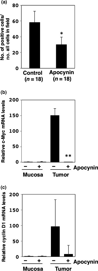
Figure 1.

Changes in cell cycle‐related factors in intestinal tumors treated with or without apocynin. (a) Immunohistochemistry was performed for determination of proliferating cell nuclear antigen (PCNA)‐positive cell numbers in tumor sections (n = 18) of small intestines of Min mice treated with 500 mg/L apocynin (n = 7) and untreated controls (n = 8). Ratio of the number of PCNA‐positive cells per whole cell in field (100 ×) is shown. Data are represented by mean ± SD. *P < 0.05 versus untreated control. Real‐time PCR analysis was carried out to obtain c‐Myc (b) and cyclin D1 (c) mRNA levels. Values were set at 1.0 in untreated controls, and relative levels were expressed as mean ± SD (n = 4, a pair of mucosa and tumor samples for apocynin or untreated controls). **P < 0.01 versus untreated control. GAPDH mRNA levels were used to normalize data.
