
Fig. 3.
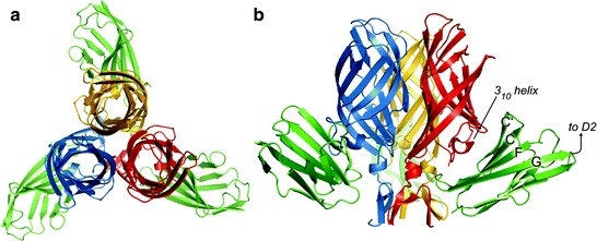
Crystal structure of the σ1-JAM-A complex. (a and b) Ribbon drawings of a complex formed between the trimeric σ1 head domain and monomeric JAM-A D1, viewed along the threefold symmetry axis (a) and from the side (b). Monomers of the σ1 head are shown in blue, red, and yellow; JAM-A D1 is shown in green. Secondary structure elements are labeled. Figure and legend modified from Kirchner et al. (2008)