
Fig. 8.
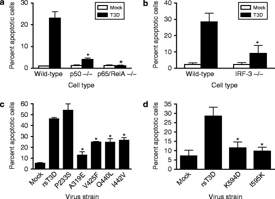
Reovirus entry triggers apoptosis dependent on NF-κB and IRF-3. (a and b) Wild-type cells or cells lacking NF-κB p50, NF-κB p65/RelA, or IRF-3 were either mock-infected or infected with T3D at an MOI of 100 PFU/cell. After incubation at 37°C for 48 h, cells were stained with acridine orange. The results are expressed as the mean percentage of cells undergoing apoptosis for three independent experiments. Error bars indicate SD. *, P < 0.05 as determined by Student’s t-test in comparison to T3D-infected wild-type cells. Figure modified from Connolly et al. (2000) and Holm et al. (2007). (c and d) HeLa cells were infected with rsT3D or each μ1 δ (c) or μ1 φ (d) mutant at an MOI of 100 PFU/cell. Following 48 h incubation, the percentage of apoptotic cells was determined by staining with acridine orange. Results are expressed as the mean percentage of apoptotic cells for triplicate samples. Error bars indicate SD. *, P < 0.05 as determined by Student’s t-test in comparison to rsT3D. Figure modified from Danthi et al. (2008a, b)