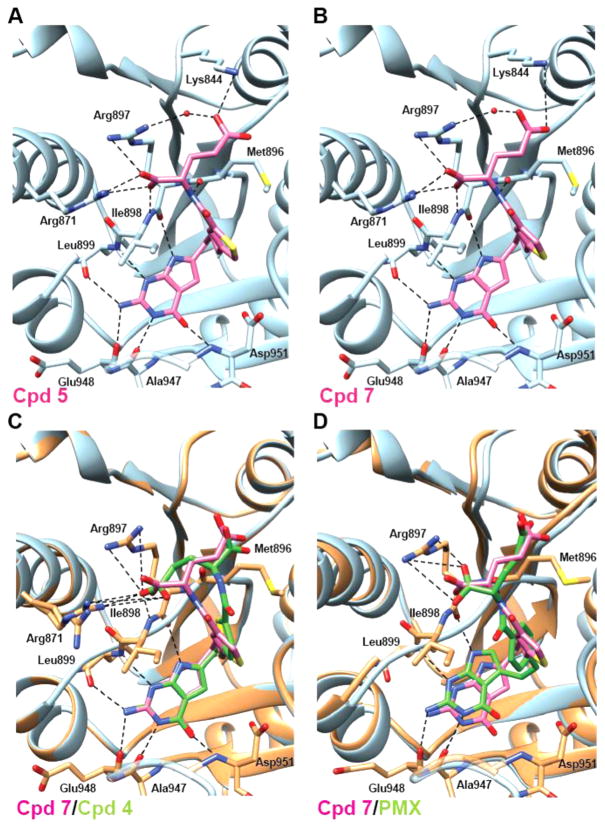
Figure 11.
Structural analyses of 5, 7, and PMX conformations in ternary complexes with human GARFTase and β-GAR. Crystal structures of the most potent GARFTase inhibitors, 5 (A) and 7 (B), are shown alongside overlays of 7 (pink) with 4 (C, green) or the less potent PMX (D, green) in the 10-formyl tetrahydrofolate binding pocket of human GARFTase with interacting residues from GARFTase shown in stick representation. (C,D) GARFTase ribbon is shown in blue for the 7 complex and in dark gray for either the 4 or PMX complex, with contacts to the latter indicated with dashed lines. Details including interaction distances and ligand electron density maps for 5, 7, and PMX complexes are included in in Table 4S and Figure 5S, Supporting Information.

