FIGURE 4.

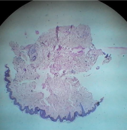
Histopathological examination (high power field, hematoxylin-eosin stain) of the lipoatrophic area shows loss of subcutaneous fat and absence of inflammatory cells in case 2.

Histopathological examination (high power field, hematoxylin-eosin stain) of the lipoatrophic area shows loss of subcutaneous fat and absence of inflammatory cells in case 2.