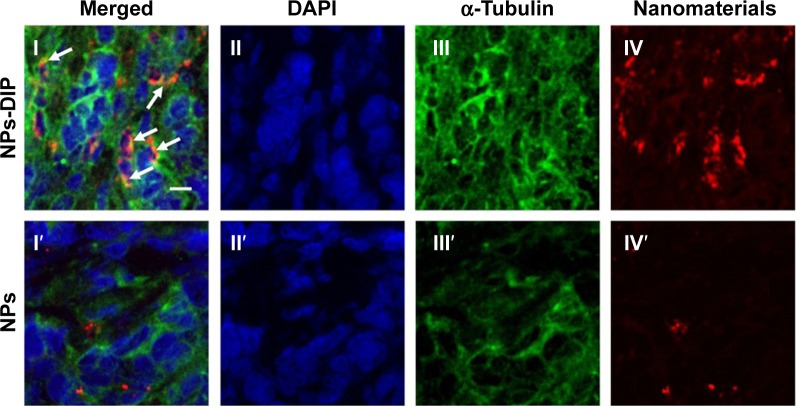
Figure 6.
Localization of nanoparticles within pancreatic tumor xenograft.
Notes: The frozen AsPC-1 tumor slices were obtained from the tumor samples (Figure 5C) and then experienced to typical IHC analysis. (I) Arrows were used to emphasize that many nanomaterials were colocalized with tubulin, indicating NPs-DIP were efficiently internalized into tumor cells. (I, I′) merged field; (II, II′) nuclei were labeled in blue with DAPI; (III, III′) cytoskeleton α-tubulin was stained in green with Alexa Fluor 488 anti-tubulin antibody; (IV, IV′) nanomaterials exhibited in red. Scale bar: 10 µm.
Abbreviations: IHC, immunohistochemistry; NPs, nanoparticles; NPs-DIP, Ser–Glu-functionalized NPs.

