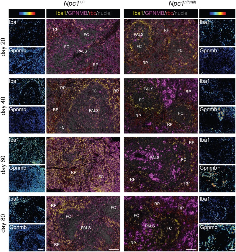
Fig 5. Gpnmb positive cells are increased with disease progression in Npc1nih/nih mice spleen.
"Composite" panels show immunostaining of Gpnmb in magenta and Iba-1 in yellow and methylgreen nuclear counterstain of spleen of Npc1+/+ and Npc1nih/nih at 20, 40, 60 and 80 days of age. Brightfield scans were analysed using spectral imaging, Iba1 and Gpnmb are displayed with a color-coded intensity scale. Red blood cells (rbc) were imaged as well (red colour, “composite” panels). FC = follicle centre, PALS = peri-arteriolar lymphocyte sheath, RP = red pulp. Scale bar = 100 μm.

