
Figure 1.
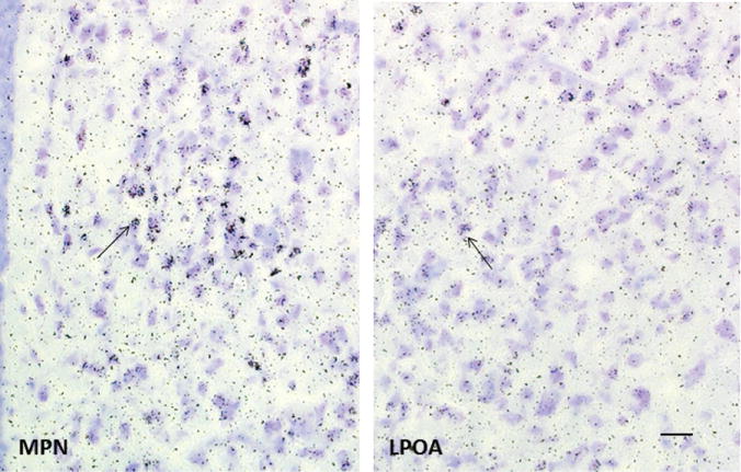
Female rats were anesthetized with pentobarbital and injection (i.v.) with 100 μCi/100 g body weight of 2,4,6,7,16,17-[3H]estradiol (specific activity 130 Ci/mmol, New England Nuclear). At 2 h after injection of the isotope, animals were killed, brains removed and cryosectioned (10 μm) through the medial preoptic nucleus (MPN) and lateral preoptic area (LPOA). Sections were collected under safe-light conditions and thaw-mounted onto slides which had been coated with nuclear track emulsion and exposed at −70°C for 28 weeks, then photodeveloped. The MPN contains many cells that concentrate 3H-estradiol as indicated by the accumulation of silver grains over nuclei (arrow). In the LPOA, some cells have accumulations of silver grains (arrow) but levels are much lower in both the estradiol-accumulating cells and the surrounding background compared with the MPN. Bar in LPOA is 20 μm and applies to both images. Sections are adapted, with permission, from a study by Akesson TR and Micevych PE (4).