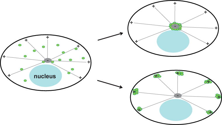
Figure 2. Application of the assay in cells with a microtubule-organizing center.

This schematic shows the predicted change in vesicle distribution resulting from an interaction between the candidate protein and labeled vesicles in the assay described in Protocol 1. Before adding linker drug, vesicles are distributed throughout the cell (left). If the candidate protein binds the vesicle, then in cells expressing FKBP-tagged Bicaudal D2, adding the linker drug results in dynein-mediated transport towards microtubule minus-ends, causing the accumulation of vesicles in the center of the cell (top right). In cells expressing FKBP-tagged KIF5C motor domain, addition of the linker drug causes accumulation of labeled vesicles in the periphery of the cell, near the plus-ends of microtubules (bottom right). If the candidate protein does not bind the labeled vesicles, then there will be no change in their distribution. Adapted from Bentley et al., 2015.
