
Figure 4. Detecting protein-vesicle interactions based on an equilibrium change in vesicle distribution.
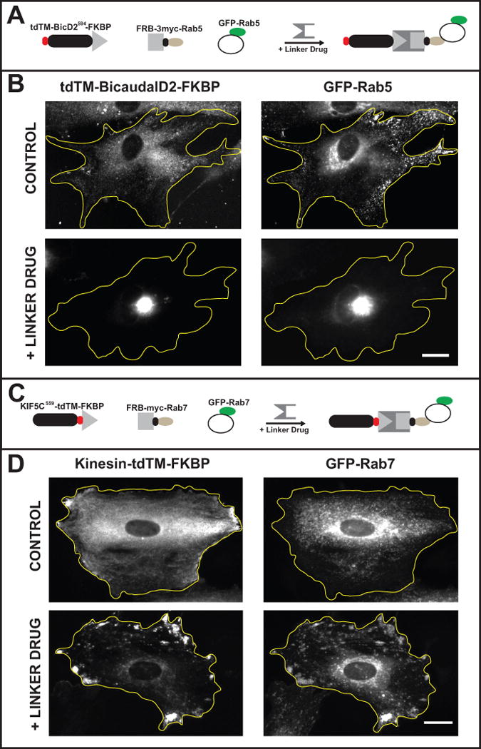
In this assay active motor proteins are targeted to labeled vesicles if, and only if, these vesicles bind the expressed FRB-tagged candidate protein. The examples show rat embryonic fibroblasts in which endosomes were mislocalized to the cell center by dynein or to the cell periphery by kinesin. (A & B) Expressing FRB-3myc-Rab5 and tdTM-BicD2594-FKBP resulted in a redistribution of early endosomes when linker drug was present. (A) A schematic showing the constructs expressed: tdTM-BicD2594-FKBP, FRB-3myc-Rab5, and GFP-Rab5. (B) Representative images show the distribution of tdTM-BicD2594-FKBP and GFP-Rab5 in control cells and in cells treated with linker drug. In control cells tdTM-BicD2594-FKBP was diffusely distributed with some vesicle association. GFP-Rab5 vesicles were distributed throughout the cell. In cells treated with linker drug, both GFP-Rab5 vesicles and tdTM-BicD2594-FKBP became concentrated in the center of the cell. (C & D) Expressing FRB-3myc-Rab7 and KIF5C559-tdTM-FKBP resulted in redistribution of late endosomes when linker drug was present. (C) A schematic showing the constructs expressed: KIF5C559-tdTM-FKBP, FRB-3myc-Rab7, and GFP-Rab7. (D) Representative images show the distribution of KIF5C559-tdTM-FKBP and GFP-Rab7 in control cells and in cells treated with linker drug. In control cells, the kinesin motor domain was diffusely distributed with small accumulations at a few points in the periphery of the cell. GFP-Rab7 vesicles had a mostly perinuclear distribution. In cells treated with linker drug, GFP-Rab7 vesicles accumulated in the periphery of the cell, together with the kinesin motor domain. The yellow lines outline cell boundaries. Bar, 30 μm. Adapted from Bentley et al., 2015.