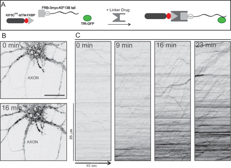
Figure 5. Detecting protein-vesicle interactions in living neurons based on directing vesicles to the axon.

In the example shown, the assay was used to identify one of the kinesins that bind to dendritic vesicles labeled with transferrin receptor-GFP. (A) A schematic showing the three constructs expressed in this assay before and after assembly of the split kinesin. (B) Images showing the cell body and proximal axon of a neuron imaged immediately before (0 min) and 16 min after addition of the linker drug. Note the increase in intensity of TfR-GFP in the axon after 16 min. Bar, 20 μm. (C) Kymographs showing the transport of transferrin receptor vesicles in the axon before and at varying times after adding the linker drug. Time is shown on the x axis and position along the axon on the y axis. Diagonal lines with positive slope represent movements away from the cell body. Before adding the linker drug there was nearly no anterograde transport of vesicles in the axon. Addition of the linker drug resulted in a pronounced increase in long-range anterograde vesicle transport in the axon. In these images the contrast was reversed so that bright vesicles appear black. Adapted from Jenkins et al., 2012.
