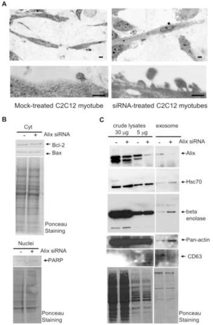
Figure 3. Alix-depletion affects release and protein content of muscle-derived nanovesicles.
A) Light microscopy analysis (top panel) and transmission electron microscopy (bottom panel) of 3D cultures of differentiated C2C12 cells (DIII) silenced for Alix showed an altered phenotype.
B) Cytoplasmic (top) and nuclear (bottom) fractions from Mock- and Alix-silenced myotubes were immunoblotted for the indicated apoptotic markers. Ponceau staining is shown as a control of the total protein loaded per lane.
C) Treatment of C2C12 myoblast with Alix-specific double stranded siRNA pools led to a significant reduction (≈80%) in Alix expression compared to mock-transfected cells. Nanovesicles released by Alix-silenced C2C12 cells exhibited a significant accumulation of Hsc70, enolase and actin, and a reduction of CD63.

