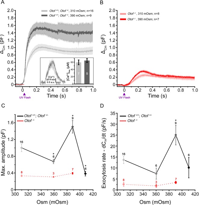
Figure 4. Exocytosis triggered by Ca2+ uncaging is sensitive to intracellular hydrostatic pressure.
(A) Exocytosis in IHCs expressing otoferlin was largely potentiated when rising osmotic pressure from 310 mOsm (light grey) to 390 mOsm (dark grey). The darker line in each condition indicates the mean responses and light filled area the standard error. The jump in the concentration of intracellular free Ca2+ was similar in 310 and 390 mOsm conditions (inset; p = 0.6). (B) Exocytosis evoked in Otof-/- IHCs in conditions similar to A. (C-D) Comparative maximum amplitude and peak exocytotic rate at various intracellular osmotic pressure. The results showed a maximum facilitation at 390 mOsm. These pressure effects were not seen on the residual slow exocytosis of Otof -/- IHCs. Data points are means ± SEM. The number of IHCs is indicated above each point. * p < 0.05.

