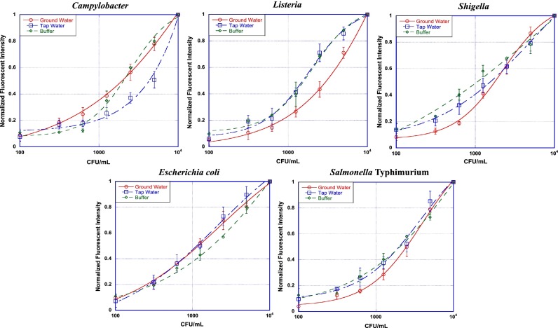
FIG. 2.
Bacterial detection in assay buffer (n = 3), tap water (n = 9), and ground water (n = 9). For detection in buffer, each bacteria in ten-fold serial dilution was incubated with its corresponding capture (5 min) and detection (15 min) antibody and spun at 8000 RPM for 1 min through density gradient. For detection in tap and ground water, 20 ml of tap or ground water were spiked with bacteria and concentrated using VivaSpin columns. Corresponding buffers were added for each bacteria and the assay were performed as explained earlier. The fluorescent signal was quantified using fluorescent microscopy. The normalized fluorescence for negative control sample is represented by concentration 100 CFU/ml.

