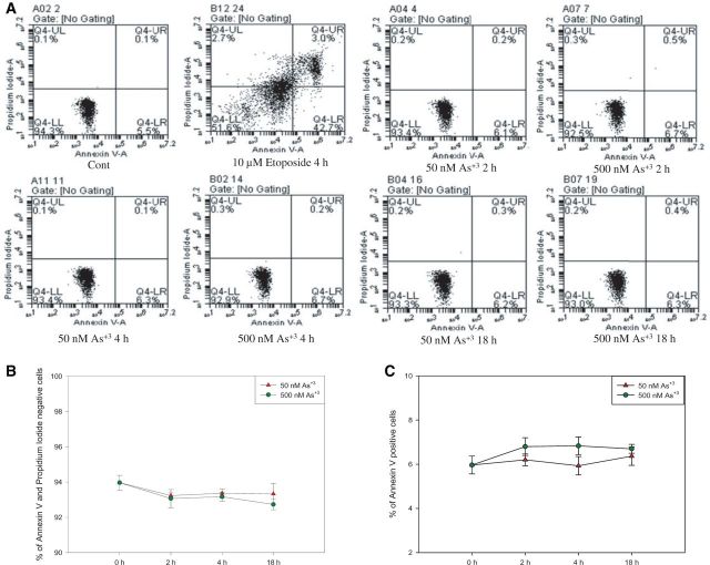
FIG. 4.
Annexin V and PI staining in D1 cells exposed to As+3. D1 cells were treated with 50 or 500 nM As+3 for 2, 4, and 18 h, or 10 µM Etoposide as positive control. A, Flow cytometry results showing D1 cells which are Annexin V-PI- (LL), Annexin V+PI- (LR), Annexin V-PI+ (UL), or Annexin V+PI+ (UR). B, Viability (% of Annexin V-PI- cells). C, % of apoptotic cells (% of Annexin V+ cells).

