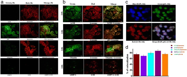
Fig. 12.

Co-localization of proteins associated with autophagy. Immunofluorescence (IF) demonstrates (a) colocalization of LC3B (green) with Lysosome (red), p62 (green) with Lysosome (red), Ubiquitin (green) with Lysosome (red), LAMP-1(green) with LC3B (red) and p62 (green) with LC3B (red) in HT-29 cells without 2c treatment. b colocalization of LC3B (green) with Lysosome (red), p62 (green) with Lysosome (red), Ubiquitin (green) with Lysosome (red), LAMP-1(green) with LC3B (red) and (c) p62 (green) with LC3B (red) counterstained with DAPI (blue) in HT-29 cells treated with 2c for 24 h. d percentage of cellular puncta co-localized for five pair of protein sets were determined. Scale bars, 10 mm. The bars represent Mean ± SEM
