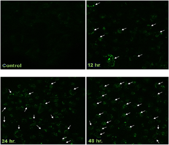
Fig. 3.

2c induced vacuolization and formation of MDC-labeled vesicles in HT-29 cells. Cells were incubated in RPMI 1640 medium. After 2c treatment with indicated time intervals, both treated and control cells (0 h) were incubated with MDC at 0.05 mM for 10 min at 37 °C followed by washing with PBS (four times) and immediately analyzed under fluorescence microscopy where the nature of the vacuoles was confirmed to be authophagic (40× magnification) with increasing intensity with respect to different time periods
