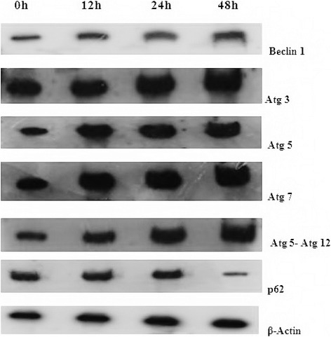
Fig. 4.

Expression of Autophagy proteins in 2c induced HT-29 cells. Cells were treated with 2c (14.9 μM for 12, 24, 48 h) and expression levels of Beclin-1, Atg 3, Atg 5, Atg 7, Atg 5-Atg 12, p62 were quantified by western blot analysis from cell lysates of control and treated cells. Analysis was confirmed with three different sets of experiments. β-actin served as a loading control
