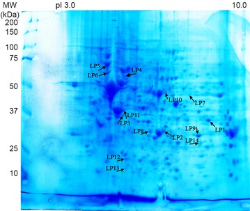
Fig. 4.

Coomassie-blue stained gel of CCLP1 2-D resolved proteins. Proteins immunoreactive with more than 30 % of the 13 CC sera are highlighted by arrows. These immunoreactive spots are listed in Additional file 1: Table S1. Isoforms of annexin A2 were recognized by 69 % of CC sera and corresponded to spots LP9 and LP14. HSP-β1 (54 % of sera) corresponded to LP12. Isoforms of annexin A1 and actin were recognized by 46 % of CC sera and corresponding spots were LP2 and LP8 (for annexin A1) and LP3 and LP11 (for actin). Fructose-biphosphate aldolase A (LP1), lamin-B2 (LP4), 78 kDa glucose-regulated protein (LP5) and isoform 2 of serine hydroymethyltransferase (LP7) were identified by 38 % of CC sera. Each of the remaining three spots were stained by only four (31 %) different sera: glutathione S-transferase (LP13), retinal dehydrogenase 1 (LP10) and vimentin (LP6)
