Fig. 2.

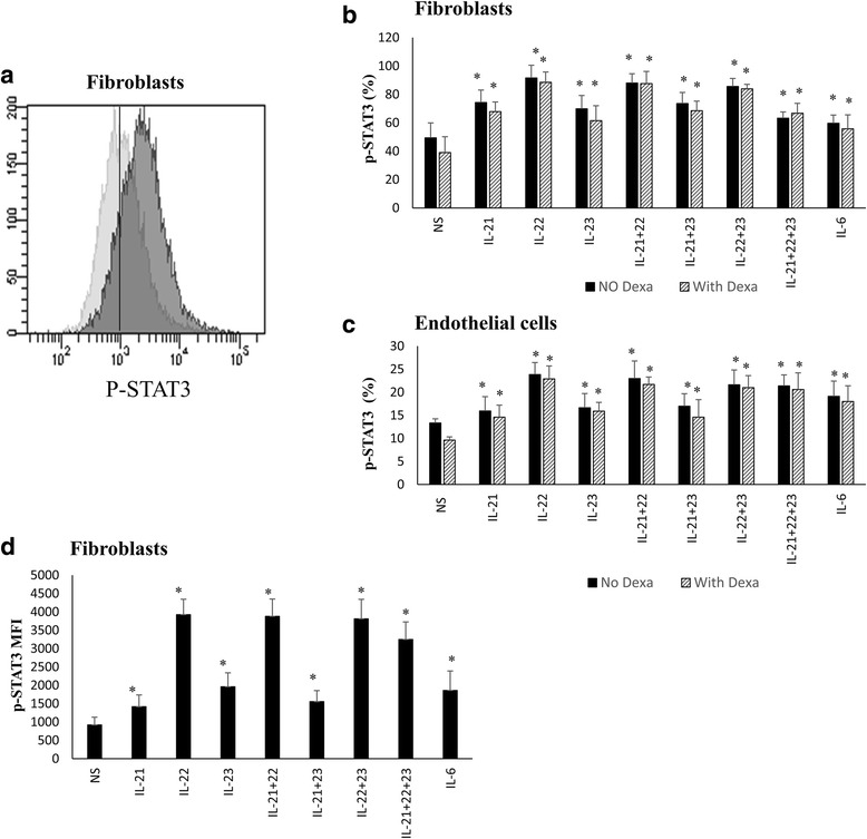
Th-17 regulatory cytokines induce STAT3 phosphorylation in fibroblasts and endothelial cells. Primary human lung fibroblasts and HMVEC-L endothelial cells were treated or not with dexamethasone (5 μM) for 1 h then stimulated with cytokines for 15 min, fixed in 4 % PFA and ice-cold methanol and stained with PE labeled-anti-p-STAT3 antibody and analysed using the BD LSRII flow cytometer. a Representative FACS data showing level of STAT3 phosphorylation in fibroblasts following IL-21+22+23 cytokines stimulation. b, c Percentage of p-STAT3 following treatment, or not, of fibroblasts (b) and endothelial cells (c) with cytokines alone or in combinations. d Mean Fluorescent Intensity (MFI) of p-STAT3 within fibroblasts following treatment with cytokines. n = 8 for each cell type. Comparison is always between cells treated with cytokines (in the presence or absence of Dexamethasone) and non-treated cells. Data is expressed as means ± SE *p ≤0.05. NS non-stimulated
