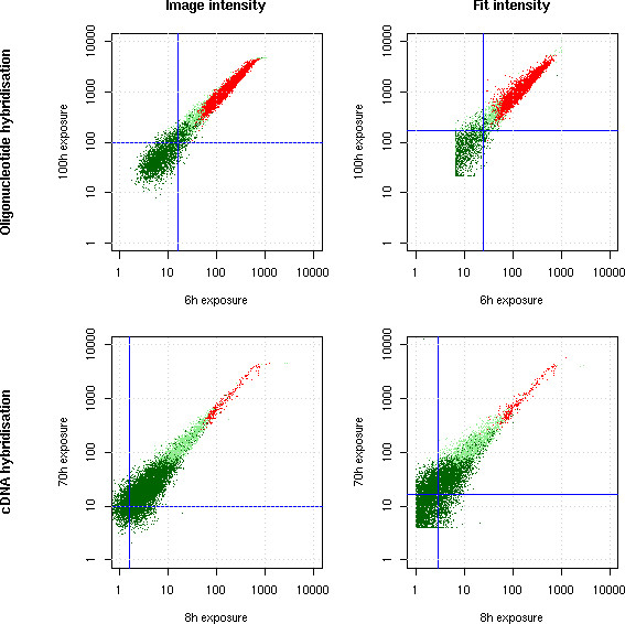
Figure 5.

Saturation correction. Two hybridisations were scanned after two different exposure times each (6 h against 100 h for an oligonucleotide hybridisation and 8 h against 70 h for a cDNA hybridisation). The image quantification mode saturates while the fit quantification preserves a linear increase in signal. The red points are detected as present in both experiments, the light green in one of the experiments and the dark green in neither (see Image Processing section for a description of this flag). Blue lines indicate background levels (median background computed by the fit equation (1). Notice that saturated spots are detected as absent in the longer exposures because of their flat top.
