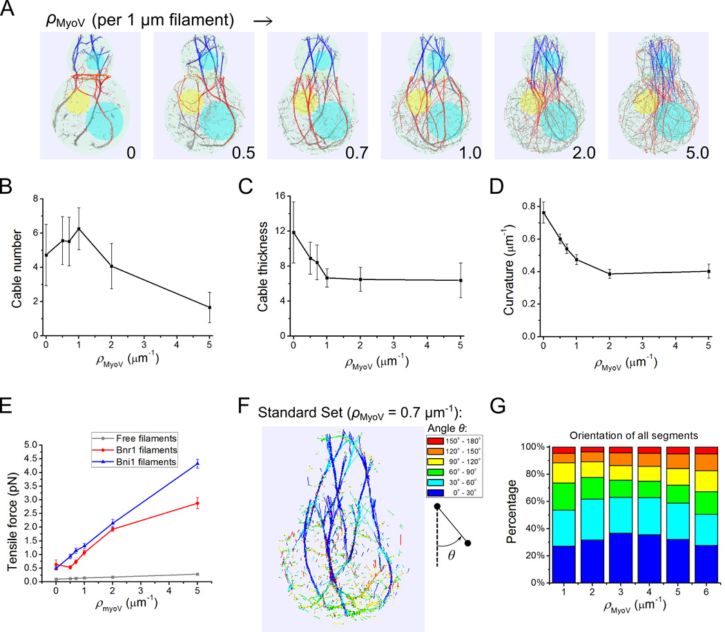
Figure 6. Myosin V pulling stretches actin filaments and influences bundling into cables in simulations.
(A) Snapshots at 60 s with increasing myosin V density ρMyoV (Movie S5). Filaments become unbundled and straighter along with ρMyoV increase. (B–E) Effects of myosin V pulling on cable number (B), thickness (C), average curvature (D) and tensile forces of Bni1, Bnr1 and free filaments segments (E). The number of cables goes through a maximum while filaments get stiffer, thinner and more tense as the myosin V density increases. All error bars are standard deviations from 15 time points (separated by 10 s starting at 60 s) of 3 independent runs. (F) Snapshot with standard parameter set (Table I, ρMyoV = 0.7 pN/µm) with segments colored according to their barbed-to-pointed end orientation with respect to the z axis. (G) Orientation distribution as a function of myosin V density ρMyoV. Legends of angle distribution are shown corresponding to the color codes in F. The degree of filament alignment goes through a maximum, similar to the cable number in panel B. Data: average from 15 time points (separated by 10 s starting at 60 s) of 3 independent runs.

