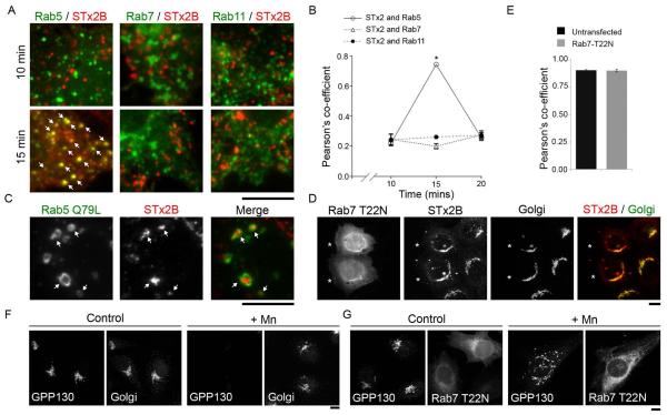
Figure 2.
Endosome-to-Golgi transport of STx2B avoids transit through Rab7-positive late endosomes.
A. Cells were transfected with indicated GFP-tagged RabWT constructs. One day after transfection, transport of His-tagged STx2BWT was assayed as described in Methods. Cultures were fixed at 10, 15 or 20 min after initiation of transport; images for the 10 and 15 min time-points are depicted here and all time-points are quantified in Panel B. After fixation, cells were stained with an antibody against the His-tag and imaged to detect GFP and STx2B. Arrows show overlap of STx2B with Rab5. Scale bar, 10 μm.
B. Quantification of the Pearson’s co-efficient for colocalization between STx2B and Rab5, Rab7 or Rab11 from Panel A (mean ± SE; n=10 cells per time-point; * p<0.05 for the difference between STx2B-Rab5 colocalization at 15 min compared to all other conditions by one-way ANOVA and Dunnett’s post hoc test).
C. Cells were transfected with GFP-tagged Rab5Q79L. One day after transfection, transport of His-tagged STx2BWT was assayed. Cultures were fixed 15 min after initiation of transport, stained with an antibody against the His-tag and imaged to detect GFP and STx2B. Arrows show presence of STx2B in Rab5Q79L-positive endosomes. Scale bar, 10 μm.
D. Cells were transfected with GFP-tagged dominant negative Rab7 (Rab7T22N). After 24 h, transport of His-STx2BWT was assayed. Cultures were fixed 60 min after initiation of toxin transport and processed to image STx2B, using an anti-His antibody; the Golgi, using an anti-GPP130 antibody; and GFP. Asterisks denote transfected cells. Scale bar, 10 μm.
E. Pearson’s co-efficient for colocalization between STx2B and the Golgi from Panel D (mean ± SE; n=10 cells per group).
F and G. HeLa cells that did not over-express globotriaosylceramide were either left untransfected (Panel F) or transfected with GFP-tagged Rab7T22N (Panel G). After 24 h, cultures were treated with or without 500 μM Mn for 4 h, fixed and imaged to detect GPP130 and the Golgi, using an antibody against SPCA1, a Golgi-localized Ca/Mn transporter (Panel F) or GFP and GPP130 (Panel G). In Rab7T22N-expressing cells, after Mn-treatment, GPP130 persists in cytoplasmic punctae, as described by us previously (22). Scale bars, 10 μm.

