
Figure 3.
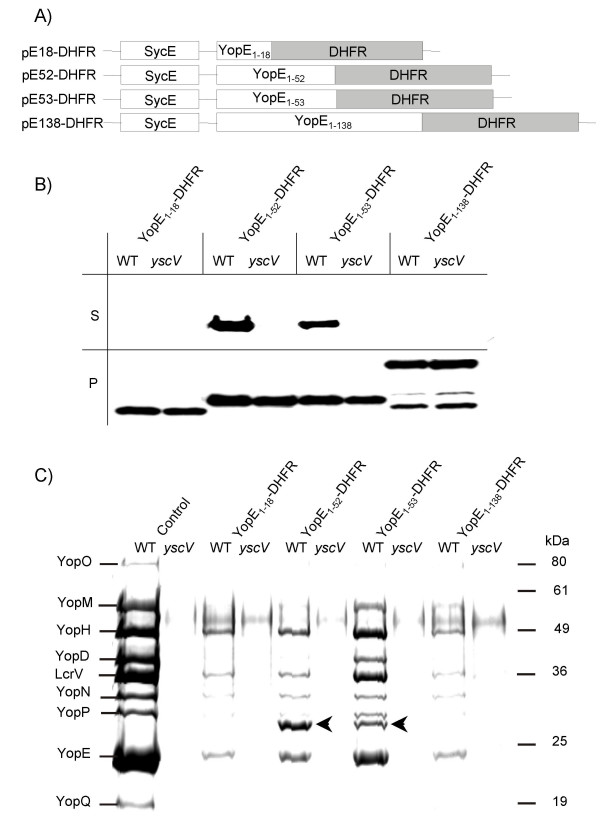
Expression and secretion of YopE-DHFR fusions. (A) Scheme depicts plasmid constructs pE18-DHFR, pE52-DHFR, pE53-DHFR and pE138-DHFR, allowing expression of YopE N-termini (the first 18, 52, 53 or 138 aa) fused to DHFR and concomitant expression of YopE-chaperone SycE. (B) Yersiniae (Y. enterocolitica strain WA-314, abbrev. WT, and its secretion deficient yscV mutant (also termed lcrD)) expressing YopE-DHFR fusions were cultured for 2 h at 37°C in BHI medium. Then Yop secretion was induced by Ca2+-depletion with EGTA. After 1.5 h of continued incubation bacteria were pelleted and whole cell lysates (P) and TCA-precipitated supernatants (S) were subjected to SDS-PAGE; ten times more supernatant than cell pellet was loaded. Subsequently, immunoblotting was performed with monoclonal anti-DHFR antibodies. (C) Coomassie-stained SDS-PAGE of supernatants from a secretion experiment as described above (Fig. 3B). The first two lanes show Yop secretion of the parental hosts. Arrows indicate secreted YopE1–52-DHFR and YopE1–53-DHFR.