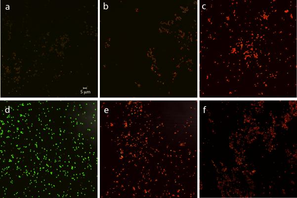
Fig. 5a-f.
BacLight staining of dormant and germinated spores with or without prior scCO2-PAA treatment or autoclaving. Spores of strain PS533 (wild-type) either untreated, autoclaved (30 min; 250°F) or treated with scCO2-PAA wet and inactivated ~ 90% or treated dry and inactivated ~ 99% were stained with BacLight and photographed by fluorescence microscopy as described in Methods. scCO2-PAA treated and untreated spores were also heat activated for 60 min at 75°C, and germinated for 45 min at 37°C in LB medium plus 10 mmol l−1 L-valine, stained with BacLight and photographed by fluorescence microscopy. The various panels in the figure are: a) dormant untreated spores; b) wet dormant scCO2-PAA treated spores; c) dormant autoclaved spores; d) germinated untreated spores; e) germinated spores that had been treated with scCO2-PAA as wet spores; and f) germinated spores that had been treated with scCO2-PAA as dry spores. The scale bar in the panel is five microns and all panels have the same magnification.

