
Figure 3.
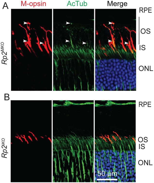
Mouse retinas from 2 months old Rp2MKO (A) or Rp2iko (B) were stained with anti-M-opsin (red) antibody or anti-acetylated tubulin (AcTub; green). Arrowheads indicate the elongated OS (depicted by black bar) in cones, as depicted by co-localized signal in merged images. RPE, retinal pigment epithelium; OS, outer segment; IS, inner segment; ONL, outer nuclear layer. Scale bar: 50 μm.