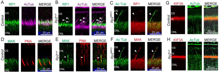
Figure 4.

Retinas from control, Rp2null and Rp2MKO mice were stained with anti- RP1 (A, B, green and C, red), MAK (D, E, green; F, red) or KIF3A (G, H, red) antibodies. Anti-acetylated α-tubulin (AcTub) antibody was used as axoneme marker. Arrowheads indicate the presence of RP1-specific and MAK-specific signal in the elongated cone outer segment of mutant retinas. G, H: Immunostaining of control and Rp2null mouse retina sections did not show KIF3A positive signal in elongated COS. Nuclei are stained with Hoechst (blue).
