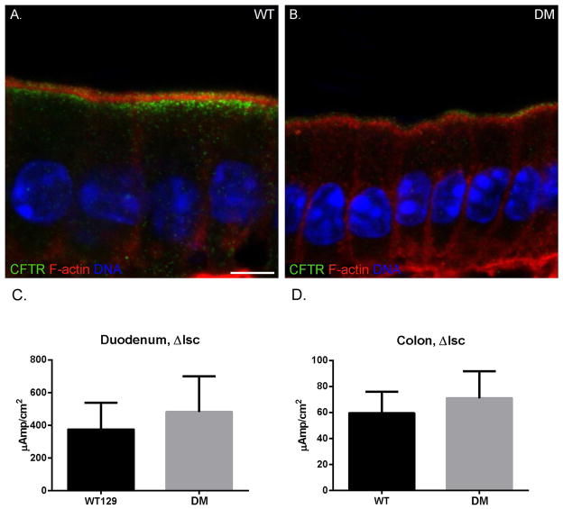
Fig. 12. The BB MV localization and cAMP-stimulated anion transport of CFTR of the DM are comparable to that of WT, but subapical endosomal expression/localization is reduced.
Localization of actin (red) and CFTR (green) in WT (A) and DM (B) duodenum. DAPI stained nuclei are shown in blue. Total CFTR staining intensity is greatly reduced in the DM due to loss of subapical localization. Bar: 5 μm. (C, D) No significant differences in the increase in isc (μamps/cm2) after forskolin stimulation in (WT n=2) and DM (n=2) duodenum (C; P=0.49) and colon (D; P=0.42) were observed.

