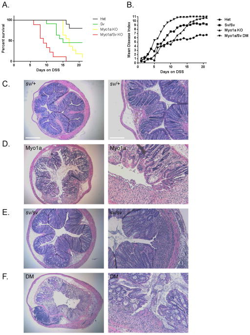
Fig. 13. Myo1a KO and sv/sv mice exhibit intermediate and DM mice high levels of hypersensitivity to DSS induced colonic mucosal injury.
(A). Survival index of sv/+ (black), sv/sv (yellow) , Myo1a KO (green) and DM (red) mice as a function of days on drinking water with 3% DSS. (B) Disease index scores as a function of days on DSS. (C–F) H&E stained sections of most affected regions of colon from sv/+ and sv/sv mice at the 21 day end point for treatment (C, E), or from Myo1a KO (D) and DM (F) mice that reached the 20% weight loss end point (for the images of mice colons shown, day 15 for the Myo1a KO and day 13 for the DM). Note that in comparison to the sv/+ control, mucosal injury, edema and submucosal immune cell infiltration is greater in all three myosin mutants. Left bar B–F: 375 μm; Right bar (B–F): 150 μm

