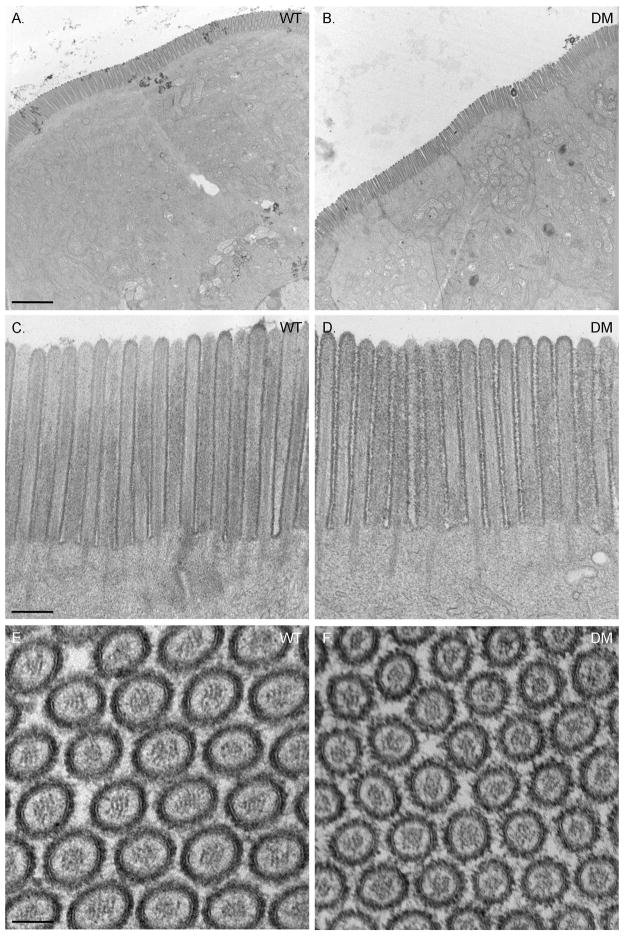
Fig. 2. The ultrastructure of the DM intestinal epithelial cell and its apical BB is WT-like in organization.
(A) Low magnification TEMs of WT and DM intestinal epithelium. (B) Ultrastructure of the WT and DM BB region. (C) Cross section through BB MV in WT and DM. Bars (A): 2 μm (B): 250 nm (C): 75 nm

