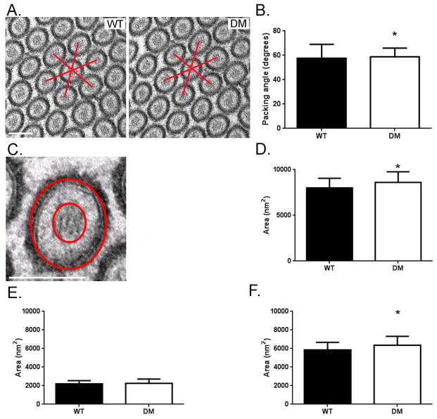
Fig. 4. MV hexagonal packing and intramembrane space between the MV membrane and MV core is restored in the DM.
(A) TEM cross sectional views of MV in WT and DM BB. Bar: B) Histogram of measured angles between adjacent WT and DM MV measured from multiple (n=606 angle measurements per genotype) cross sectional TEM images. Both WT and DM MV exhibit hexagonal packing with DM exhibiting even tighter packing than WT (WT, 57.7° ± 11.3°; DM, 58.7° ± 7.2°; *p < 0.05). C) High magnification cross sectional view of a DM MV showing the MV core, inter-MV membrane-core space and MV membrane. MV and MV core cross sectional areas were determined by measuring the area within the outer and inner circles respectively; intra-MV free space area was determined by subtracting the inner circle area from the outer circle area. Bar: 100 nm D) Histogram of mean MV cross sectional area shows that DM microvilli have slightly increased diameter (~102 nm vs. 105 nm) as compared to WT (WT, 8025 nm2 ± 1033 nm2, n = 46 vs. DM, 8596 nm2 ± 1171nm2, n = 46 ; *p < 0.005). E) Histogram of mean MV actin core area shows that there is no statistical difference between WT and DM (WT, 2198 nm2 ± 836 nm2, n = 46 vs. DM, 2269 nm2 ± 445 nm2, n = 46). F) Histogram of mean MV free space area between plasma membrane and actin core shows that there is slightly increased free space in the DM as compared to WT consistent with the increase and total MV diameter and comparable MV core diameter (WT, 5826 nm2 ± 836 nm2, n = 46 vs. DM, 6327 nm2 ± 998 nm2, n = 46; *p < 0.005).

