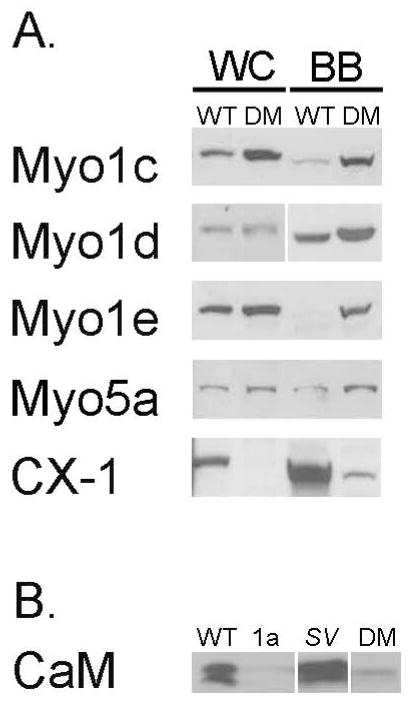
Fig. 5. Multiple myosins are recruited to the DM BB.
(A) Immunoblot analysis of isolated WT and DM BBs for Myo1c, d, e and Myo5a indicate that these myosins exhibit increased expression/association in the isolated DM BB. Total class I myosin expression, based on immunoblot with the monoclonal antibody CX-1, is still less than in WT BB. (B) Comparison of calmodulin content in isolated BBs from WT, sv/sv, Myo1a KO and DM. The 20-17 kDa doublet bands seen in the WT and sv/sv lanes reflect the differential migration of apo- and Ca2+-calmodulin (Chacko et al. 1994).

