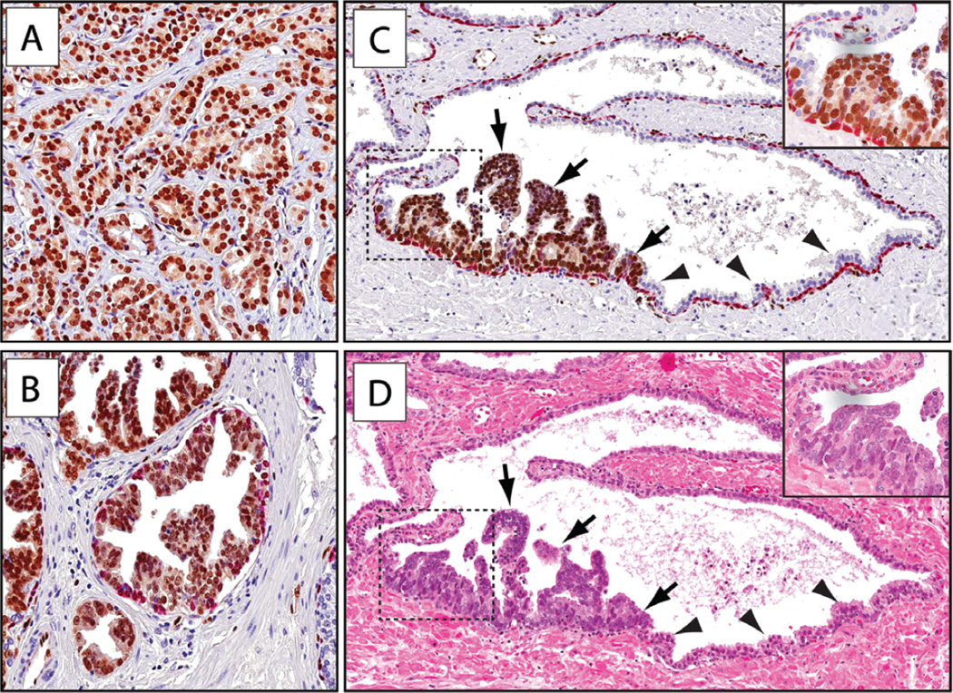
Figure 1.
ERG protein expression in carcinoma and HGPIN. Sections were subjected to immunohistochemical staining for ERG (brown) and basal cell-specific markers (p63/903, red) (original objective magnification 20×). (A) Representative micrograph of ERG-expressing tumour. (B) HGPIN (tufting type) lesion showing ERG expression. (C) Partial ERG-positive PIN lesion with ERG-expressing neoplastic cells (arrows) adjacent to normal prostatic epithelium (arrowheads). (D) H&E stain of adjacent section showing distinct HGPIN morphology of the ERG-positive cells.

