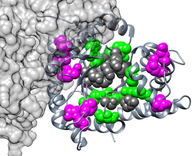
Fig. 3. Top EVC Positions for Aldolase.

This view of aldolase is looking into the active site of one monomer. The top 20 consensus EVC positions (35, 43, 47, 53, 56, 106, 107, 122, 145, 148, 168, 169, 193, 214, 234, 237, 267, 270, 275, and 300; magenta and green spacefilled) of tetrameric human aldolase C are shown on one monomer (PDB: 1xfb58). Active site positions are highlighted in dark gray. EVC positions in contact with active site residues are highlighted in green; those without contact are highlighted in magenta.
