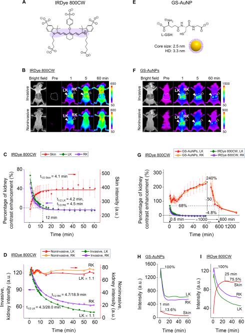
Figure 2.
Comparison of renal clearable NIR emitting IRDye 800CW and GS-AuNPs in noninvasive kidney imaging. (A) Chemical structure of IRDye 800CW. (B) Whole-body invasive and noninvasive fluorescence images of mice before and after iv injection of IRDye 800CW (Ex/Em filters: 710/790 nm). The skin-removed areas were marked by dashed white line. LK, left kidney, RK, right kidney. (C) Time-course changes in skin intensity and percentage of kidney-contrast enhancements of mice after iv Injection of IRDye 800CW. N = 3, mean ± s.d. (D) Time-fluorescence intensity curves (TFICs) of kidneys obtained from noninvasive and invasive fluorescence imaging of mice after iv injection of IRDye 800CW. (E) Schematic representation of GS-AuNPs. (F) Whole-body invasive and noninvasive fluorescence images of mice before and after iv injection of GS-AuNPs (Ex/Em filters: 710/830 nm). (G) Comparison of time-dependent kidney contrast enhancements in percentage after GS-AuNPs and IRDye 800CW injection, respectively. N = 3, mean ± s.d. (H,I) TFICs of kidneys and skin obtained from invasive imaging after iv injection of GS-AuNPs (H) and IRDye 800CW (I).

