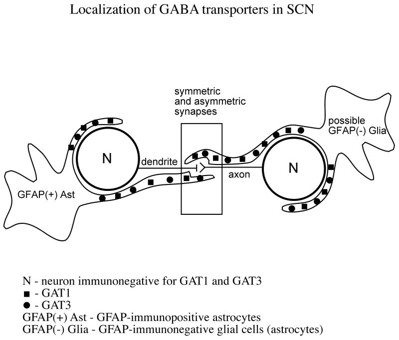
Figure 11. Schematic model of the location of GATs in SCN.
This model schematically proposes that GAT1 and GAT3 are expressed in the distal processes of GFAP-positive astrocytes, and also by GFAP-negative glial cells. The model shows that GATs-ir processes of glial cells surround neuronal cell bodies and axo-dendritic synapses. The location of the GATs suggests they can restrict GABA diffusion from the synapses and regulate extracellular GABA concentration around the neuronal cell bodies.

