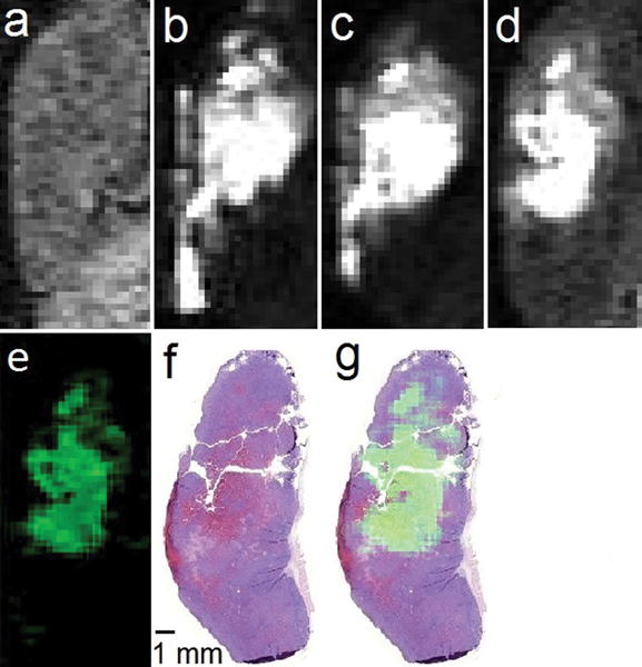
Figure 3.

T1-weighted in vivo sagittal plane images of a 4T1 tumor injected with EuII-222Fb (a) pre-injection; (b) 3 min, (c) 20 min, and (d) 120 min post-intratumoral injection; (e) difference between the 120 min and pre-injection images (image d minus image a) colored using the ImageJ green lookup table; (f) hematoxylin- and eosin-stained slice of tumor imaged in a–e; and (g) sum of images e and f. All images are on the same scale. Imaging parameters included an echo time of 1.5 ms, repetition time of 11 ms, flip angle of 40°, field of view of 30 mm × 90 mm, and an in-plane resolution of 0.352 mm × 0.352 mm.
