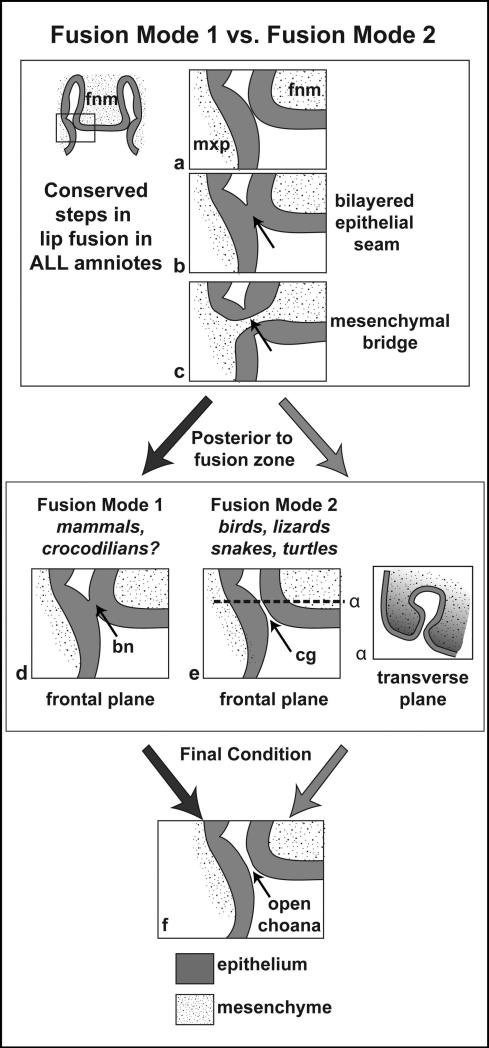
Figure 4.
Schematic summarizing the two modes of primary palate fusion. Steps a - c depict processes in primary palate fusion that are conserved in all amniotes, although the actual prominences which initiate the fusion varies according to taxon. Steps d and e depict Fusion Modes 1 and 2 respectively. In step d, just posterior to the mesenchymal bridge unifying the primary palate, the epithelial seam (nasal fin) persists, forming a transient bucconasal membrane. In e, there is no epithelial seam posterior to the primary palate; instead entering the choanal groove. In the transverse plane (α), the decline in proliferation at the base of the choana (lighter grey) allows for the groove to remain as the prominences grow around it. The final state (f) in all amniotes is in an open choana connecting the oral and nasal cavities. bn, bucconasal membrane; cg, choanal groove. Figure modified from (Abramyan et al., 2015).

