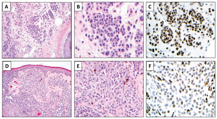
Fig. 1.
Representative photomicrographs showing 5-hydroxymethycytosine (5-hmC) immunolabeling in a melanocytic nevus (A–C), markedly decreased 5-hmC expression in a primary melanoma (D–F). 5-hmC nuclear labeling is also observed in background normal cells including keratinocytes and lymphocytes (not shown). Panels A and D at ×100; B, C, E, and F at 400×, original magnification.

