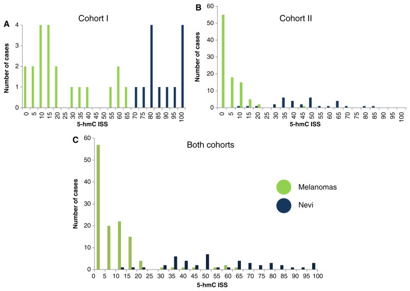
Fig. 4.
Histogram showing the distribution of immunohistochemical staining scores for 5-hydroxymethycytosine (5-hmC) expression for melanocytic nevi and melanoma. An immunohistochemical staining score of zero (0) indicates no 5-hmC immunolabeling in any of the evaluable lesional nuclei, where an immunohistochemical staining score of 100 indicates that all lesional nuclei were markedly/strongly immunoreactive (see Methods for detailed description for calculating immunohistochemical staining score). When the two cohorts were evaluated separately, (A) and (B) respectively, the potential discriminatory ability of 5-hmC immunohistochemistry between nevi and melanoma is more evident than when both cohorts are combined (C).

