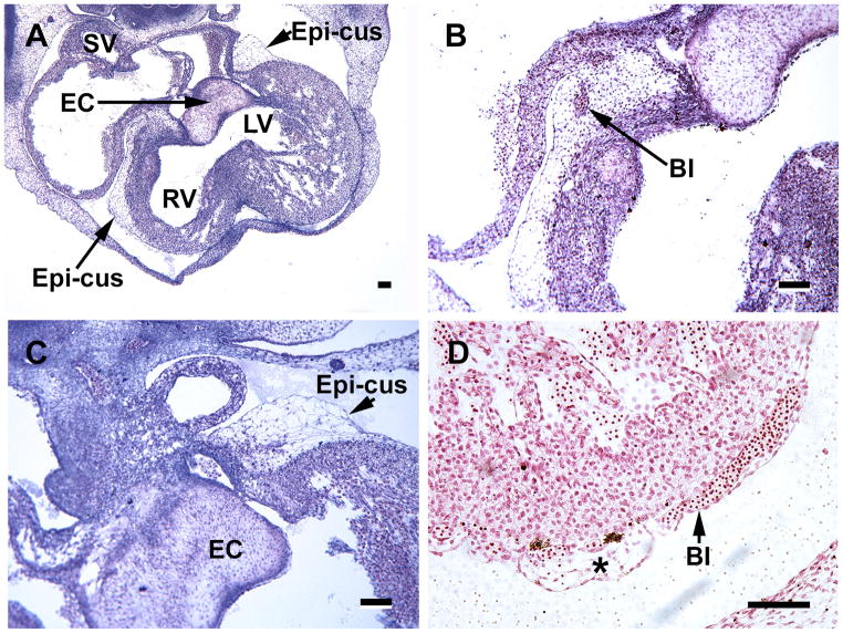
Figure 2.
Hearts from S17 embryos illustrating the importance of the mesenchymal-containing epicardial atrioventricular cushions (Epi-cus), at the atrioventricular groove. These structures represent the expanded subepicardium. A, an overview of the heart indicates that they are present at both right (RV) and left (LV) ventricles near the endocardial cushions (EC); the sinus venosus (SV) is also seen in this image. B, a blood island (BI) appears within the epicardial cushion. C, the endocardial cushion and the mesenchyme-filled epicardial cushion are seen at a higher power than in A. D, subepicardial blood islands (BI) are seen in close association with epicardial-derived subepicardial cells (*). Magnification bar = 100 μm.

