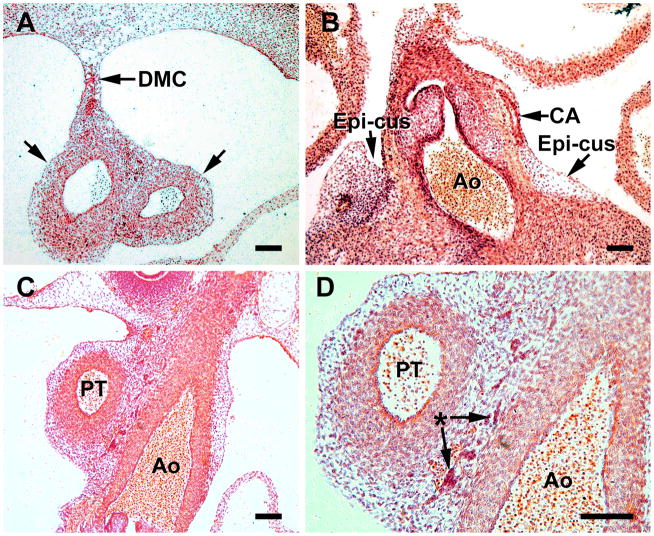
Figure 4.
S18 heart sections comprise this figure, which represents the time of coronary ostia and stem formation. A, In this transverse section at the level of the arterial trunk, the dorsal mesocardium (DMC) is seen attaching to the aorta and pulmonary trunk (between arrows) and serves as a source of cells for the arterial trunk. B, formation of a coronary artery (CA) passing through the persisting epicardial cushion (epi-cus) at the aortic root (Ao). C, a network of cells (mostly mesenchymal) surrounds the pulmonary trunk (PA) and aorta (Ao). D, a higher power of C. The asterisk indicates blood islands and isolated erythrocytes that are characteristic of this region. Magnification bar = 100 μm. B,C and D are from coronal sections.

