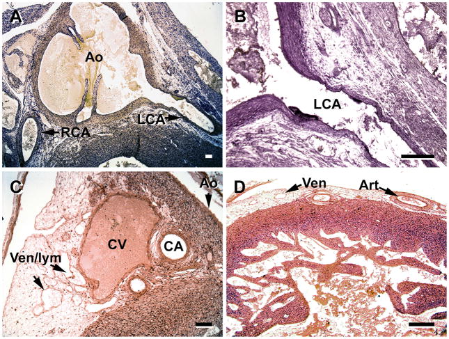
Figure 8.
A and B, by the 12th week the coronary arteries (CA) of fetal hearts have a well-developed tunica media. C and D, the venous drainage has expanded as seen by numerous venules, although some of the structures may be lymphatic vessels (Ven/lym ). C, great cardiac vein (CV) at the base of the aorta (Ao) has a complete tunica media, and the coronary artery branches (CA) have well-developed meda. D, the number of veins and arteries (in the left ventricle) have increased substantially in the subepicardium. Ven/lym, veins/lymphatic vessels; RCA, right coronary artery, LCA, left coronary artery. Slides in A and B were stained with Masson trichrome. Magnification bar = 100 μm.

