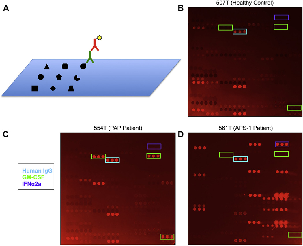
FIG 1.
Protein microarrays for ACAA detection. A, Schematic representation of cytokine microarrays. Cytokines, chemokines, growth factors, and traditional autoantigens are printed onto a nitrocellulose-coated microscope slide. Arrays are probed with serum (green) and detected by using fluorophore-conjugated secondary antibody (red). B–D, Representative images of protein microarrays probed with serum from a healthy control subject (Fig 1, B), a patient with PAP (Fig 1, C), and a patient with APS-1 (Fig 1, D). A control IgG feature printed on the array is boxed in cyan, GM-CSF features are boxed in green, and IFN-α2a features are boxed in purple.

