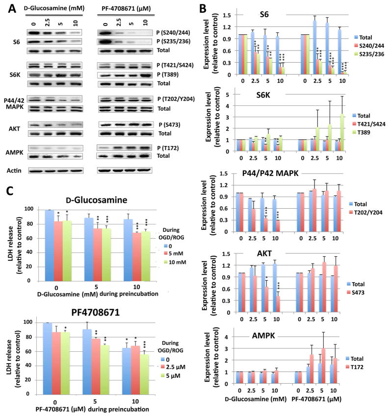
Figure 8. Effect of the S6K inhibitors, D-glucosamine and PF-4708671, on phosphorylation of S6K-rpS6 signaling proteins, and OGD/ROG-induced cell death in rat cortical neurons.
(A) Rat cortical neurons were incubated with glucosamine at 0, 2.5, 5 and 10 mM (left panels) or with PF-4708671 at 0, 2.5, 5, 10 μM (right panels) for 16 h; total cell lysates were analyzed for the total and phosphorylation levels of rpS6, S6K, p44/42MAPK, AKT and AMPK by Western blot. Actin was used for loading controls. (B) Quantification of protein (total and/or phosphorylated) levels. Densities of each protein band(s) were measured, normalized with corresponding actin levels and shown as relative to control (no drugs). Data are shown as the mean ± SD of three independent experiments. *P<0.05, **P<0.01, ***P<0.001 compared to control by the student’s-test. (C) Upper panel: cells were preincubated without, with 5 mM or 10 mM D-glucosamine for 16 h, and challenged to OGD (5h) followed by ROG (16 h) in the absence or presence of 5 or 10 mM D-glucosamine. Lower panel: cells were preincubated without, with 2.5 μM, or 5 μM PF-4708671 for 16 h, and challenged to OGD/ROG in the absence, or presence of 2.5 or 5 μM PF-4708671. Cell death was assessed by measuring the released LDH at the end of the procedure. Results are displayed in relation to the amount of released LDH in the control sample (no exposure of the drug neither during preincubation nor during OGD/ROG) (i.e. the first column in each figure). Data are shown as the average of four (D-glucosamine) or three (PF-4708671) independent experiments with standard deviations. *p<0.05, **p<0.01, ***p<0.001 by the student’s t-test compared to the control (no exposure to the drug at all, the first lane of each graph).

