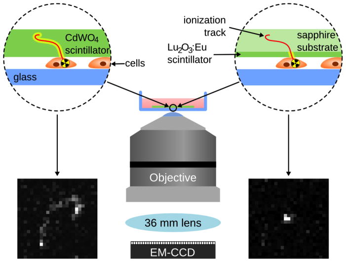
Figure 1.
Schematic of a typical radioluminescence microscopy setup using a 500 μm CdWO4 scintillator (left) and a 10 μm Lu2O3:Eu scintillator (right). The thin-film Lu2O3:Eu scintillator produces a truncated ionization track as shown here, as compared to the thicker CdWO4 scintillator, which produces a longer ionization track.

