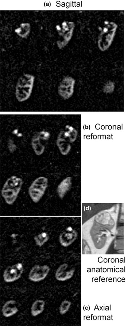
Figure 3.

Three dimensional perfusion difference images in Patient 2 initially acquired in the sagittal plane with near-isotropic resolution 2.6×2.6×2.8 mm (a) allowed for reformatted images in b) coronal, and c) axial orientations, displayed with 11.2 mm slice thickness. Perfusion is clearly high compared to surrounding parenchyma and of a heterogeneous nature, which correlates well with the anatomical appearance of the lesion (2D multi-slice single-shot FSE) shown in d). Scan time for this 3D image data was ~5 min.
