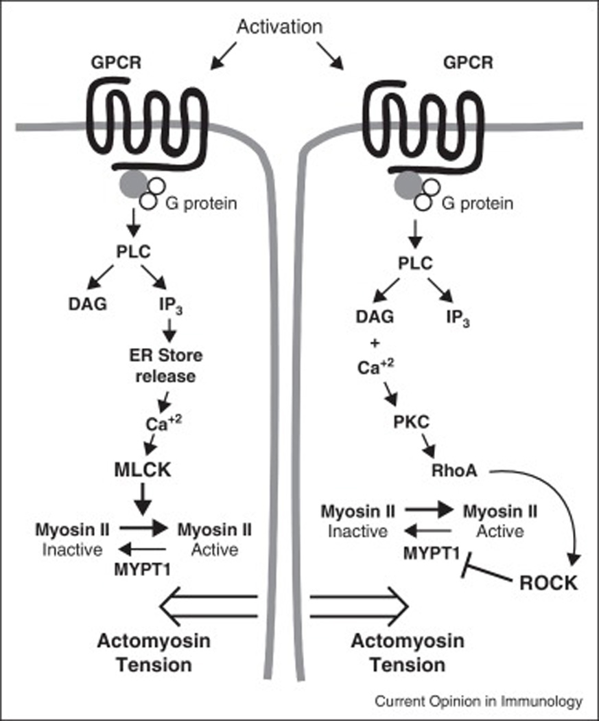
Figure 1. Global signals open junctions for vascular permeability.

The diagram depicts stimulation of G protein coupled receptors (GPCR) by a prototype soluble agonist. The activation of phospholipase C (PLC) results in the conversion of phosphatidyl inositol in the plasma membrane to the second messengers diacyl glycerol (DAG) and inositol triphosphate (IP3). (Left pathway) IP3 stimulates calcium release from stores in the endoplasmic reticulum. The calcium activates myosin light chain kinase (MLCK) to phosphorylate myosin light chain promoting activation of myosin II and actomyosin tension. This is believed to cause contraction of the endothelial cells weakening the junction and promoting vascular permeability. (Right pathway) In a parallel pathway DAG and calcium activate protein kinase C (PKC), which activates the GTPase RhoA, which in turn activates Rho-associated coiled coil kinase (ROCK), which phosphorylates and inactivates myosin phosphatase target subunit 1 (MYPT1) to inactivate it. Since MYPT1 serves to dephosphorylate and hence activate Myosin II light chain, this further stabilizes the actomyosin tension in the cells.
